Microfracture
When a patient suffers an injury to the cartilage surface of the knee, usually the femur, it can cause a defect in the cartilage that will never grow back. This area of cartilage loss is at high risk of developing osteoarthritis due to the loss of covering of the underlying bone. Detached cartilage around the major defect can also cause problems with trapping in the joint causing locking.
Treatment of such choral defects is difficult. There are some specialist techniques that attempt to repair the defect with new cartilage cells under flaps of synovial tissue, or by treatment with Stem cells, which POTENTIALLY can form new cartilage cells. However, neither of these techniques has strong evidence of success.
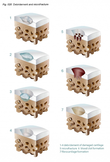
A technique called MICROFRACTURE involves drilling into the hard bone under the cartilage at the base of the defect. It is performed as part of an arthroscopic (keyhole) surgical technique. This allows bone marrow and blood clot (haematoma) to collect in the defect. This is then allowed to stabilise and heal. The combination of bone marrow and haematoma slowly forms a scar over the defect. Although not true articular (or joint) cartilage, the scar covers the defect and creates a smoother surface where the defect once was. Micro fracture is ideal for defects of 2cm sq or less and has good evidence of successful outcomes.